Article

Exploring the Antibiotic Resistance Profile of Clinical Klebsiella pneumoniae Isolates in Portugal

1
INIAV—National Institute for Agrarian and Veterinarian Research, Rua dos Lagidos, 4485-655 Vila do Conde, Portugal
2
LEPABE—Laboratory for Process Engineering, Environment, Biotechnology and Energy, Faculty of Engineering, University of Porto, Rua Dr. Roberto Frias, 4200-465 Porto, Portugal
3
AliCE—Associate Laboratory in Chemical Engineering, Faculty of Engineering, University of Porto, Rua Dr. Roberto Frias, 4200-465 Porto, Portugal
4
Center of Biological Engineering (CEB), Campus de Gualtar, University of Minho, 4710-057 Braga, Portugal
5
LABBELS—Associate Laboratory, Braga/Guimarães, 4710-057 Braga, Portugal
6
Department of Veterinary Sciences, Laboratory Medical Microbiology, Antimicrobials, Biocides and Biofilms Unit, University of Trás-os-Montes and Alto Douro, 5000-801 Vila Real, Portugal
7
CITAB—Center for the Research and Technology Agro-Environmental and Biological Sciences, University of Trás-os-Montes and Alto Douro, 5000-801 Vila Real, Portugal
8
Inov4Agro—Associate Laboratory for Innovation, Capacity Building and Sustainability in Agri-Food Production, University of Trás-os-Montes and Alto Douro, 5000-801 Vila Real, Portugal
*
Author to whom correspondence should be addressed.
Antibiotics 2022, 11(11), 1613; https://doi.org/10.3390/antibiotics11111613
Submission received: 13 October 2022 / Revised: 7 November 2022 / Accepted: 9 November 2022 / Published: 13 November 2022

Abstract

:
While antibiotic resistance is rising to dangerously high levels, resistance mechanisms are spreading globally among diverse bacterial species. The emergence of antibiotic-resistant Klebsiella pneumoniae, mainly due to the production of antibiotic-inactivating enzymes, is currently responsible for most treatment failures, threatening the effectiveness of classes of antibiotics used for decades. This study assessed the presence of genetic determinants of β-lactam resistance in 102 multi-drug resistant (MDR) K. pneumoniae isolates from patients admitted to two central hospitals in northern Portugal from 2010 to 2020. Antimicrobial susceptibility testing revealed a high rate (>90%) of resistance to most β-lactam antibiotics, except for carbapenems and cephamycins, which showed antimicrobial susceptibility rates in the range of 23.5–34.3% and 40.2–68.6%, respectively. A diverse pool of β-lactam resistance genetic determinants, including carbapenemases- (i.e., blaKPC-like and blaOXA-48-like), extended-spectrum β-lactamases (ESBL; i.e., blaTEM-like, blaCTX-M-like and blaSHV-like), and AmpC β-lactamases-coding genes (i.e., blaCMY-2-like and blaDHA-like) were found in most K. pneumoniae isolates. blaKPC-like (72.5%) and ESBL genes (37.3–74.5%) were the most detected, with approximately 80% of K. pneumoniae isolates presenting two or more resistance genes. As the optimal treatment of β-lactamase-producing K. pneumoniae infections remains problematic, the high co-occurrence of multiple β-lactam resistance genes must be seen as a serious warning of the problem of antimicrobial resistance.

1. Introduction

Klebsiella pneumoniae is a gram-negative opportunistic pathogen capable of colonizing, invading, and causing infections in diverse anatomical sites of the human body [1,2]. Unfortunately, over the last decades, the indiscriminate use of antibiotics in human health and agriculture has contributed to the emergence of the antibiotic resistance problem [3]. Consequently, the growing resistance of K. pneumoniae infections has become a burden for clinicians. β-lactam antibiotics, including penicillin derivatives, cephalosporins, monobactams, and carbapenems, are the most widely used class of drugs in the treatment of bacterial infections [4]. For this reason, the uncontrolled emergence of resistance mechanisms against antibiotics in this family is particularly frightening. Several mechanisms of antibiotic inactivation used by different microorganisms have already been identified, but the production of antibiotic-inactivating enzymes is the most common mechanism [5]. In the case of β-lactams, these enzymes are called β-lactamases, including carbapenemases (CPN), extended-spectrum β-lactamases (ESBL), and Ambler class C cephalosporinases (AmpC) [6].
Resistance to carbapenems is a major concern in human health. CPN may confer resistance to virtually all β-lactams, a reason why this class of β-lactam is considered a highly effective antibiotics in the treatment of serious and life-threatening bacterial infections [7]. They are poorly inhibited by classical β-lactam inhibitors, except for a subgroup called metallo-β-lactamases (MBLs) [8]. It is important to note that K. pneumoniae is the most reported carbapenemase-producing bacteria, while the KPC-type carbapenemase, an Ambler class A β-lactamase, is the most frequently associated [8,9]. Other relevant CPN enzymes are MBLs, Ambler class B β-lactamases that include Imipenem-resistant Pseudomonas (IMP)-carbapenemases and Verona integron-encoded metallo-β-lactamase (VIM) types, New Delhi metallo-β-lactamase type 1 (NDM-1), and the class D carbapenemases known as OXA-type β-lactamases [8,10].
ESBLs are a group of plasmid-mediated enzymes belonging to Ambler class A of β-lactamases, which can hydrolyze most penicillins, monobactams, and cephalosporins, and hydrolyze third- and fourth-generation cephalosporins classified as critically important antimicrobials. However, they are not able to degrade cephamycins and carbapenems and are inhibited by β-lactamase inhibitors (e.g., clavulanic acid, sulbactam, and tazobactam) and diazabicyclooctanones (e.g., avibactam) [11,12]. From a clinical point of view, the predominant families of ESBL are CTX-M-like, TEM-like, and SHV-like [13].
AmpC-type cephalosporinases are Ambler class C β-lactamases that can hydrolyze penicillins, monobactams, and first-, second-, and third-generation cephalosporins (cephamycins as well as to oxyimino-β-lactams), but neither fourth-generation cephalosporins (e.g., cefepime) or carbapenems. Furthermore, they are poorly inhibited by classical ESBL inhibitors [14]. The appearance of cephalosporinases in bacterial species lacking chromosomal AmpC genes, such as K. pneumoniae, is due to the so-called plasmid-mediated AmpC β-lactamase genes. There are several types of mobile AmpC genes identified, and the most widely disseminated are the CMY-2-like and the inducible DHA-like β-lactamases [13]. Although the acquired AmpC β-lactamase has been spread widely, its overall frequency has remained far below that of ESBLs [15].
Considering that the development of antibiotic resistance mechanisms requires the presence of specific resistance genes, genotypic characterization is a valuable tool to identify and/or confirm potential resistance mechanisms and provide the most effective treatment [15]. Moreover, the study of the distribution and prevalence of antibiotic resistance genes is pivotal from an epidemiological point of view in order to understand their dissemination in an increasingly connected world [16]. In this study, we first analyzed the antimicrobial susceptibility through the VITEK 2 compact system complemented with the disk diffusion method, and then, we used four multiplex PCR protocols capable of detecting the most commonly acquired β-lactamase genes of CPN (blaKPC-like, blaOXA-48-like, blaOXA-23-like, blaNDM-1-like, blaVIM-like, and blaIMP-like), ESBL (blaSHV-like, blaTEM-like, and blaCTX-M-like), and AmpC (blaCMY-2-like and blaDHA-like) families, to characterize 102 MDR K. pneumoniae isolates from patients admitted in two central hospitals in northern Portugal.

2. Results

2.1. Distribution of the K. pneumoniae Isolates

In total, 102 K. pneumoniae isolates collected by the laboratories from two Portuguese hospitals between 2010 and 2020 were used during this work. Strains were isolated from patients admitted to different areas of health care within the hospitals. Despite the source samples being diverse, the most significant was urine (39.2%), rectal swabs (15.7%), blood cultures (12.7%), and sputum (8.8%). Figure 1 shows the source of all isolates included in this work.
To facilitate further analysis, the isolates were divided into five main groups according to sample source: urine (39.2%), rectal swabs (15.7%), respiratory-associated infections (15.7%) (including sputum, bronchial aspirates, and bronchoalveolar aspirates), blood cultures (12.7%), and other samples (16.7%).

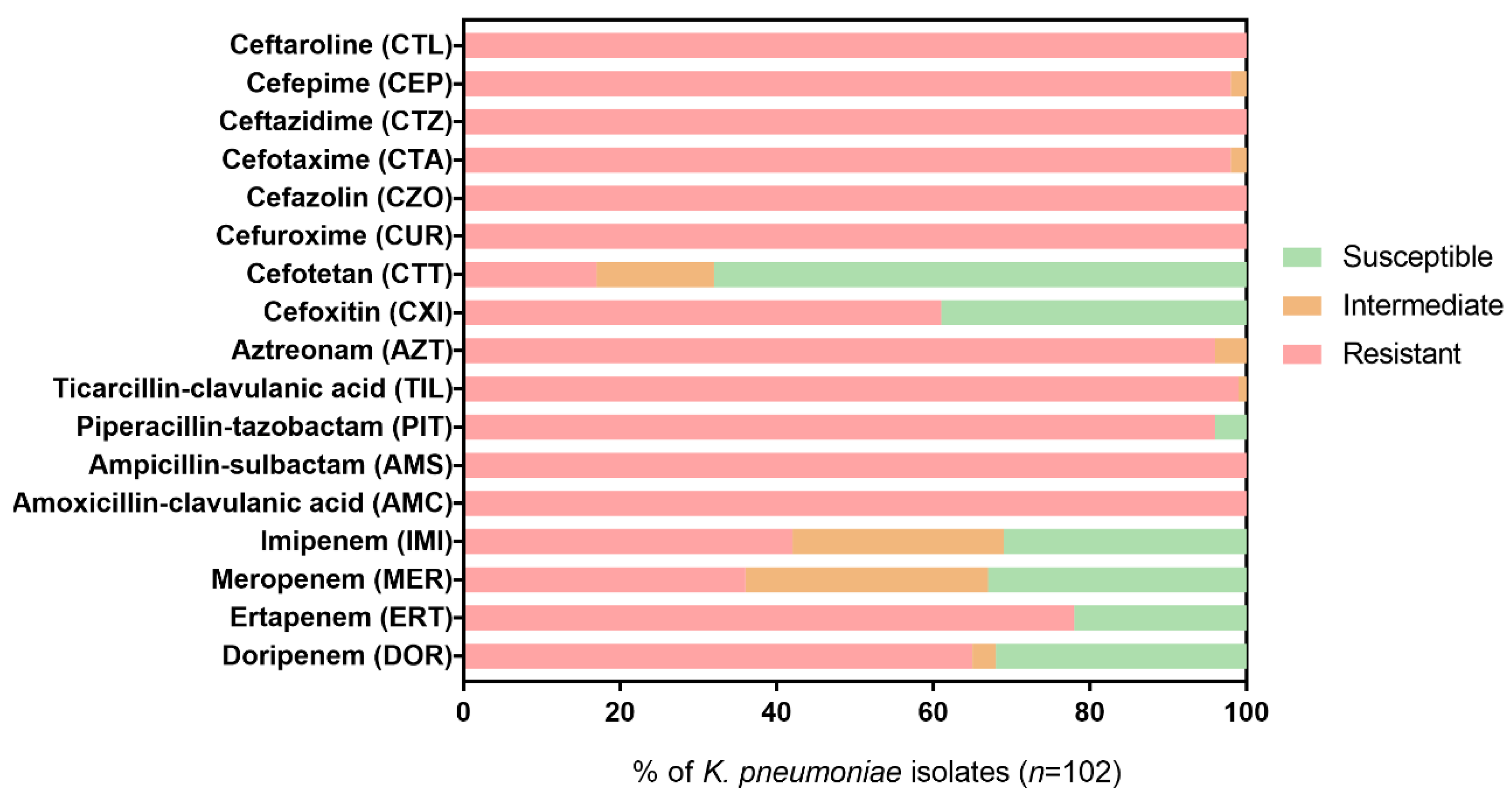
2.2. Resistance Profiles of K. pneumoniae Isolates to β-Lactam Antibiotics

The susceptibility rates of K. pneumoniae isolates for each β-lactam tested (Table 1) are shown in Figure 2. Ertapenem (ERT) was effective (i.e., susceptible profile) against 24/102 (23.5%) isolates, imipenem (IMI) was effective against 33/102 (32.4%) isolates, meropenem (MER) was effective against 35/102 (34.3%) isolates, and doripenem (DOR) was effective against 34/102 (33.3%) isolates.
The susceptibility rates of the other β-lactams were lower or even null for most isolates, with resistance rates >90% for all β-lactams tested other than the carbapenems. Only cephamycins, cefoxitin (CXI), and cefotetan (CTT) exhibited greater effectiveness (40.2% and 68.6%, respectively) against K. pneumoniae isolates. Furthermore, 21 (20.6%) K. pneumoniae isolates showed a complete non-susceptibility profile to all β-lactams tested, including carbapenems.
Based on individual antimicrobial profiles, each isolate was classified as a potential producer of CPN, ESBL, or AmpC β-lactamases according to the criteria recommended by EUCAST for the detection of resistance mechanisms and described in the “Materials and methods” (see Table S1). Notably, all K. pneumoniae isolates showed a potential ESBL-producing phenotype, while 10 (9.8%) isolates had a characteristic phenotype of both AmpC- and/or ESBL-producing bacteria. In addition, 31 (30.4%) isolates exhibited a resistance phenotype that can be classified as CPN- and/or ESBL-producing isolate, and 50 (49.0%) isolates showed a resistance phenotype that can be classified as CPN-, AmpC-, and/or ESBL-producing isolate.

2.3. Screening of Acquired β-Lactam Resistance Genes

Prior to the experiments, four multiplex PCRs were designed and optimized for the detection of the most commonly acquired β-lactam resistance genes using primers described in the literature (Table 2). Figure S1 shows representative sequences of each of the 11 bla gene families of interest with the binding site of the primer pairs used for PCR detection.
The 102 K. pneumoniae clinical isolates were then tested for the presence of the resistance genes according to the PCR methodology described in “Materials and methods”. Figure 3 shows the amplification products of the control strains separated on 1.5% (w/v) agarose gel.
The profile of resistance genes of each K. pneumoniae isolate can be found in Table S1. In total, blaKPC-like was detected in 74/102 (72.5%) K. pneumoniae isolates, while blaOXA-48-like was detected only in three isolates (2.9%). No K. pneumoniae isolates harbored blaOXA-23-like or MBL genes. For the ESBL genes, 76/102 (74.5%) were positive for the presence of blaSHV-like, 38/102 (37.3%) for the presence of blaCTX-M-like, and 47/102 (46.1%) for the presence of blaTEM-like. Among the AmpC genes, 4/102 (3.9%) isolates were positive for blaCMY-2-like, and 4/102 (3.9%) isolates were positive for blaDHA-like. blaKPC-like was most frequently detected in isolates from urine (32.4%), followed by rectal swabs (14.7%), respiratory-associated infections (8.8%), and blood (7.8%). The isolates positive for blaOXA-48-like were only associated with urine (2.0%), and respiratory (1.0%) isolates. Regarding ESBL genes, the distribution by sample sources is wider. blaTEM-like was detected in the highest percentage in urine, followed by respiratory-associated infections, other samples, blood culture, and rectal swabs. The most prevalent ESBL gene (blaSHV-like) was most associated with urine, followed by respiratory-associated infections, other samples, rectal swabs, and blood cultures. In turn, blaCTX-M-like was most detected in murine, followed by respiratory-associated infections, blood cultures, other samples, and rectal swabs. blaCMY-2-like and blaDHA-like were detected in 2.0% of urine isolates and 1.0% of respiratory-associated infections and other isolates. This data can be seen in Figure 4A.
In Figure 4B, it is possible to analyze the relative frequency of each antimicrobial resistance (AMR) gene by type of sample. Within the urine isolates (n=40), blaKPC-like was most frequent (0.83/1), followed by blaSHV-like (0.68/1), blaTEM-like (0.45/1), and blaCTX-M-like (0.43/1). In isolates from respiratory-associated infections (n=16), blaSHV-like (0.94/1) was the most prevalent, followed by blaTEM-like (0.63/1), blaKPC-like (0.56/1), and blaCTX-M-like (0.50/1). In rectal swabs (n=16), blaKPC-like was most prevalent (0.94/1), followed by blaSHV-like (0.69/1) and blaTEM-like (0.31/1. In blood cultures (n=13), blaSHV-like was most prevalent (0.69/1), followed by blaKPC-like (0.62/1), blaTEM-like (0.46/1), and blaCTX-M-like (0.46/1). Within the isolates from other samples (n=17), blaSHV-like was the most frequent (0.88/1), followed by blaKPC-like (0.56/1blaTEM-like (0.50/1) and blaCTX-M-like (0.31/1).
Only one (1.0%) isolate did not provide any positive result for all the genes evaluated, with all the other isolates having at least one acquired β-lactam resistance gene. In total, 19/102 (18.6%) revealed the presence of one, 35/102 (34.3%) revealed the presence of two, 32/102 (31.4%) revealed the presence of three, 14/102 (13.7%) revealed the presence of four, and one (1.0%) isolate revealed the presence of five acquired β-lactam resistance genes.

3. Discussion

K. pneumoniae belongs to the WHO’s list of antibiotic-resistant pathogens of critical priority that require the development of new antibiotics to combat them [24]. This alarming superbug is also part of the group of pathogens called ESKAPE (Enterococcus faecium, Staphylococcus aureus, Clostridium difficile, Acinetobacter baumannii, Pseudomonas aeruginosa, and all Enterobacteriaceae) normally described for their ability to “escape” from the general antimicrobial therapy [25]. Furthermore, K. pneumoniae is a well-known nosocomial pathogen that, in recent years, has been established as an MDR and pandrug-resistant problem [26]. Therefore, understanding the genetic determinants involved in AMR in this bacterial species is important from a clinical and epidemiological point of view [27].
The MDR K. pneumoniae isolates used in this study came from a wide range of samples (Figure 1). The main sample sources were urine (39.2%), rectal swabs (15.7%), respiratory-associated infections (15.7%; including sputum, bronchial aspirates, and bronchoalveolar aspirates), and blood (12.7%). Urine represents almost 40% of the origin of the isolates, confirming the increasing trend of MDR K. pneumoniae in urinary tract infections (UTIs) [1,28,29]. Furthermore, rectal swabs, respiratory-associated infections, and blood also represented an important source of MDR K. pneumoniae, as already reported in several other studies [1,30].
Antimicrobial susceptibility tests revealed a small variety of β-lactam treatment options for the MDR K. pneumoniae isolates from Portuguese hospitals (Figure 2). Since this study started from a pool of antibiotic-resistant clinical isolates, high-resistance rates were expected in the susceptibility tests. Therefore, the resistance percentages reported here should not be seen as the prevalence/frequency rates or compared with other studies but as molecular epidemiology data of nosocomial MDR K. pneumoniae in Portugal. Still, the high diversity of genetic determinants found in MDR K. pneumoniae isolated from two central hospitals confirms that β-lactam efficacy might be seriously threatened [31,32]. Recently, high resistance to β-lactams, including penicillins (60.5%), cephalosporins (66.9%), and carbapenems (60.5%) was also reported among a large collection of K. pneumoniae from Portuguese hospitals [33]. In other countries of the European Union/European Economic Area (EU/EEA), the AMR among K. pneumoniae isolates is also a serious problem. According to the ECDC’s annual epidemiological report, the AMR percentage for third-generation cephalosporins and carbapenems was 33.9% and 10.0%, respectively, in 2020 [34]. Furthermore, the carbapenem resistance was almost always combined with resistance to several other key antimicrobial groups, leading to a severely limited range of treatment options for K. pneumoniae. The highest percentages of carbapenem resistance (>10%) were observed in south and south-eastern European countries. In Portugal, there has been an increasing annual trend of carbapenem resistance among K. pneumoniae since 2016 (5.2%% in 2016, 8.6% in 2017, 11.7% in 2018, 10.9% in 2019, and 11.6% in 2020), exceeding the overall prevalence for Europe [34]. This trend is also observed in different studies conducted in other countries, such as China and the United States, with prevalence values reaching close to 15% or 25%, respectively [35,36]. Even so, carbapenems remain the most effective β-lactam antibiotics for most species, so they should be rationally used for the treatment of severe cases involving MDR bacteria, for which traditional β-lactams are not effective [37]. Alternately, the results suggest that cephamycins may be an alternative to carbapenems for the treatment of K. pneumoniae infections. Although cephamycins are very similar to cephalosporins and often included in the 2nd generation classification, they are resistant to degradation by ESBL enzymes, and it seems, to a certain extent, to CPN enzymes (especially CTT) [38]. However, they are reported not to be effective against AmpC cephalosporinases and porin mutations, and as noted, most cephamycin-resistant isolates in this work revealed AmpC genes [39].
The classification of potential ESBL- or AmpC-producing species was based on the resistance phenotype to both cefotaxime (CTA) and ceftazidime (CTZ), plus resistance to CXI in the AmpC-producing species. In this way, a phenotype resistant to CTA and CTZ and susceptible to CXI resulted in a classification of potential ESBL-producing bacteria. However, although a CTA, CTZ, and CXI-resistant phenotype corresponds to a potential AmpC-producing isolate, the possibility that it is also an ESBL producer cannot be discarded. Therefore, a phenotype resistant to CTA, CTZ, and CXI was classified as a potential AmpC- and/or ESBL-producing bacteria. Furthermore, it is important to note that for an isolate to be a potential CPN producer, it does not need to have an MER-resistant phenotype. According to the EUCAST criteria, a phenotypic result to MER <28 mm and PIT-resistant with disk diffusion test can be a potential CPN producer. For this reason, some K. pneumoniae isolates susceptible to MER (≥22 mm with disk diffusion) may be potential carriers of CPN genes. Indeed, 16 K. pneumoniae isolates presented an MER susceptible phenotype (Figure 2) but were classified as potential CPN-producing isolates (Supplementary Material Table S1). The classification of resistance mechanisms based only on direct evaluation of susceptibility phenotype to individual antimicrobial agents can be a simple way to categorize the resistance of clinical isolates, but it can be ambiguous. Phenotype assessment could be improved by using combinatorial testing of different antimicrobial agents to detect the specific production of β-lactamase classes, for example, by using a combination disk test, double-disk synergy test, or biochemical tests as recommended by EUCAST [15]. Still, PCR screening for acquired antibiotic resistance genes appears to be a more direct way of assessing the resistance potential of clinical isolates.
In general, ESBLs and KPC carbapenemase genes were the most detected (Figure 4), confirming their dissemination in MDR K. pneumoniae isolates as verified in several other studies [33,40,41,42,43,44,45,46]. KPC enzymes were originally identified in K. pneumoniae which justifies its high prevalence in cases of resistance to carbapenems in this species. However, this type of resistance gene has emerged in other bacterial species, such as E. coli and E. cloacae isolates [46]. In Europe, blaKPC-like (mainly KPC-2 and KPC-3) has already been detected in isolates from Austria, Belgium, the Czech Republic, Germany, Greece, Italy, Romania, and the UK, demonstrating its wide dissemination [47]. In a European survey on carbapenemase-producing Enterobacteriaceae, blaKPC (18.9%) was the major resistance mechanism reported in clinical isolates from Italy, Greece, Portugal, and Israel [48]. In other Portuguese studies, blaKPC genes were also the most detected in clinical K. pneumoniae isolates collected in the same time period of our study [33,45,49]. The highest percentage of blaKPC-like positive isolates were from urine, which confirms the high prevalence of this pathogen in UTIs [1,29]. In turn, OXA-48 is a carbapenemase that is endemic in North Africa, the Middle East, India, and some European countries such as Turkey, Spain, France, Belgium, the Czech Republic, and Malta [46,48]. However, it has also been detected in K. pneumoniae collected from Portuguese hospitals in other studies [33,45]. Recent outbreaks of OXA-48-producing K. pneumoniae in Spain, Turkey, and Greece have revealed a serious risk of these strains to human health since they present higher virulence, while laboratory detection is difficult due to their weak capacity to hydrolyze carbapenems [50,51,52]. Nonetheless, in our study, OXA-48-producing K. pneumoniae was detected in a low percentage of the K. pneumoniae isolates. Regarding MBL, no associated-resistance genes were detected in the isolates. However, the presence of blaVIM-like and blaNDM-1 genes was previously seen in K. pneumoniae clinical isolates in Portugal [53,54]. ESBL genes were the most detected in MDR K. pneumoniae isolates, demonstrating its high dissemination in Portugal, as is verified in the rest of Europe [55]. Moreover, the overall prevalence of blaSHV-like was higher than blaKPC-like. The high percentage of all ESBL types is directly related to the high percentage of resistance to β-lactams other than the carbapenems, including penicillins and cephalosporins. ESBL-producing K. pneumoniae isolates containing blaSHV-like, blaCTX-M-like, and/or blaTEM-like genes were also reported in high percentages in other clinical isolates from Portuguese hospitals, demonstrating their high spread in this bacterial species. However, blaCTX-M-like was found in a higher percentage than blaSHV-like and blaTEM-like, in accordance with epidemiology data worldwide [56]. In our study, there were more samples from around 2010 than in 2020, which is why the dominance of CTX-M-types seen in Europe may not yet be reflected in the results. In fact, the prevalence of blaCTX-M-like has been increasing considerably when compared to other ESBL types, not only in Europe but worldwide [57,58,59]. Regarding acquired AmpC β-lactamases, the low prevalence in K. pneumoniae agrees with other studies in the UK, Ireland, and Norway [60,61]. In Portugal, it was also detected among Klebsiella spp. in clinical settings, although blaDHA in a much higher percentage than blaCMY-2 [62].
The distribution by sample types was similar to all β-lactamase genes, being detected most often in urinary samples, followed by rectal swabs, respiratory-associated infections, and blood cultures. blaOXA-48-like was only detected in urinary isolates and respiratory-associated infections, whereas acquired AmpC β-lactamases, blaDHA-like and blaCMY-2-like genes, were detected in isolates from urine, respiratory-associated infections, and pus. However, it is important to notice that the number of isolates for each sample type is very different (ranging from 40 urinary isolates to 13 blood cultures), which means that for sample types with more isolates, the distribution of the genetic determinants is certainly more accurate. An analysis within each sample type revealed that blaKPC-like is highly associated with rectal swab isolates and urine isolates (Figure 4B). This high association of KPC carbapenemases in rectal swabs has already been described in several studies [63,64,65] since K. pneumoniae is an intestinal pathogen subject to a huge selective pressure by antibiotic use. On the other hand, in samples from respiratory-associated infections, blood cultures, and other samples, the frequency of ESBL genes seemed higher compared to carbapenemases genes, in most cases higher than any carbapenemase gene. For example, blaSHV-like had an almost total frequency (0.94/1), higher than blaKPC-like (0.56/1), as well as blaTEM-like (0.63/1) for isolates from respiratory-associated infections. Still, for all sample types, the frequency of ESBL genes was highest for blaSHV-like, followed by blaTEM-like, and finally, blaCTX-M-like.
Regarding the MDR K. pneumoniae isolate with no resistance genes detected, given the enormous diversity of resistance elements, this isolate might simply carry a less common resistance gene different from those tested or have another resistance mechanism (e.g., efflux pumps or porins modifications) responsible for the phenotypic pattern [66]. All other isolates tested positive for one or more resistance genes corresponding to at least one of the predicted resistance phenotypes (Table S1). Still, not all phenotypes predicted for each isolate have been confirmed by the presence of resistance genes. As mentioned above, resistance to different β-lactams can result from the production of enzymes that directly hydrolyze these or from the combination of enzymes, such as AmpC and/or ESBL, with other cellular mechanisms, such as decreased membrane permeability to antibiotics [67]. It also needs to be considered that the development of resistance depends on the level of gene expression, and thus, it cannot be excluded that resistance genes cannot express in-vitro (susceptibility tests) but show expression in-vivo and/or truncated β-lactamase genes may be detected by molecular methods but not be expressed [68]. Lastly, resistance phenotypes may result from the production of β-lactam resistance genes other than those tested in this study.
In addition, approximately 80% of MDR K. pneumoniae isolates were found to have two or more tested resistance genes (Table S1). This evidence may be associated with the high capacity for clonal expansion and exchange of mobile genetic elements (e.g., β-lactam resistance genes) of this species, which promotes increased resistance to antibiotics [67]. That said, the potential for resistance of this important nosocomial pathogen is alarming, and this work highlights the urge to introduce in the marketplace of new alternative therapies to fight infections due to K. pneumoniae.

4. Materials and Methods

4.1. Bacterial Isolates

One hundred and two K. pneumoniae isolates with an MDR profile were provided by the microbiology laboratories of the Hospital de Braga and of the Centro Hospitalar De Trás Os-Montes E Alto Douro, E.P.E., both in northern Portugal. Isolates were collected over a period of ten years (2010–2020). This study was performed in line with the principles of the Declaration of Helsinki and the guidelines of Good Clinical Practice. Approval was granted by the Ethics Committee of Life and Health Sciences Research Institute (ICVS), School of Health Sciences, University of Minho, Braga, Portugal. The isolates provided were previously identified using VITEK®2 Compact B System (bioMérieux, Marcy-l’Étoile, France). Even so, the bacterial isolates were cultured on MacConkey agar (BioLife, Milan, Italy) plates, and typical K. pneumoniae colonies were sub-cultured onto trypticase soy agar (TSA, Liofilchem, Roseto degli Abruzzi, Italy) for further analysis. Isolates were also preserved in 20% (v/v) glycerol (Riedel-de-Haën, Seelze, Germany) in tryptic soy broth (TSB, Liofilchem) and stored at −80 °C. The sample sources and isolation dates from all isolates were gathered together with the bacterial species information.

4.2. Antimicrobial Susceptibility Testing

Antibiograms were obtained by a VITEK 2 compact system available at the hospital laboratories. The antibiotic susceptibility profiles were then confirmed and complemented to meet the recommended list of antimicrobial agents proposed by the European Center for Disease Prevention and Control (ECDC) and the Centers for Disease Control and Prevention (CDC) [67], using the reference disk diffusion method as described by the EUCAST [68]. The list of β-lactam antibiotics tested is shown in Table 1. Quality control (QC) was performed using Escherichia coli ATCC 25922 and EUCAST breakpoint tables, version 12.0, were used in the interpretation of the susceptibility results [69]. Based on the antimicrobial susceptibility profiles, the K. pneumoniae isolates were classified as potential producers of CPN, ESBL, and/or AmpC according to the EUCAST guidelines for the detection of resistance mechanisms of clinical and/or public health importance [15]. In summary, the isolates were considered suspected of CPN-producing Enterobacteriaceae if the result of susceptibility to meropenem with disk diffusion was <28 mm. To be considered a potential ESBL-producing Enterobacteriaceae, the isolates required an intermediate or resistance result to one or both of cefotaxime (CTA) and ceftazidime (CTZ), while AmpC β-lactamase-producing Enterobacteriaceae required a resistant profile for CTA or CTZ, and cefoxitin (CXI).

4.3. Detection of Acquired β-Lactam Resistance Genes by Multiplex PCR

Four multiplex PCR assays for the detection of the most common β-lactam resistance genes of CPN, ESBL, or AmpC were designed using primers described in the literature. The different multiplexes were optimized using positive control strains containing the resistance genes of interest (Table 2). Subsequently, the K. pneumoniae isolates were screened for the presence of the acquired β-lactam resistance genes. In brief, a loop (1 µL) of each bacterial culture was homogenized in 500 µL of ultra-pure water and then boiled for 15 min. The suspension was centrifuged (12,000× g) for 5 min, and the supernatant (2 µL) was used as a DNA template. PCR was carried out in a 20 μL reaction mixture containing 10× reaction buffer (NzyTech, Lisbon, Portugal), 1.5 mM MgCl2, 0.25 mM of dNTPs (dATP, dTTP, dGTP, and dCTP), five pmol each of forward and reverse primers and 1.25 U of Supreme NZYTaq II DNA polymerase (NzyTech) in an MJ Mini Personal Thermal Cycler (Bio-Rad, CA, USA). The PCR conditions included initial denaturation at 95 °C for 5 min, followed by 30 cycles of denaturation at 94 °C for 30 s, annealing for 30 s at an optimized temperature, and extension at 72 °C for 30 s. A final extension was set at 72 °C for 5 min. The PCR products were mixed with 6x NZYDNA loading dye (NzyTech), subjected to electrophoresis at 100 V for one h in a 1.5 % (w/v) agarose gel, previously stained with GreenSafe Premium (NzyTech), and finally analyzed under UV light.

4.4. Statistical Analysis

All data were analyzed with GraphPad Prism version 7 (La Jolla, CA, USA) using one-way ANOVA with Holm-Sidak’s multiple comparisons test. Values with a p < 0.05 were considered statistically significant.

5. Conclusions

In summary, this work applied four multiplex PCRs to rapidly detect the presence of the most common acquired resistance genes of carbapenemases, ESBL, and AmpC β-lactamases. As a fast, reliable, and low-cost methodology, this can be a valuable tool in epidemiological studies as confirmation of standard phenotypic characterization methodologies. Overall, it was observed a diverse pool of β-lactam resistance genetic determinants disseminated among K. pneumoniae isolates (blaKPC-like, blaOXA-48-like, blaTEM-like, blaCTX-M-like, blaSHV-like, blaCMY-2-like and blaDHA-like), revealing a diverse threat to the effectiveness of the β-lactam class. In addition, this long-term study confirmed the high spread of multiple β-lactam resistance genes among K. pneumoniae isolates from hospital settings in Portugal, highlighting the need for mitigation measures to prevent the advancement of this problem.

Supplementary Materials

The following supporting information can be downloaded at: https://www.mdpi.com/article/10.3390/antibiotics11111613/s1, Table S1. Antimicrobial susceptibility profiles and genetic determinants of resistance of each K. pneumoniae isolate. Figure S1. Example sequence of each β-lactamase gene deposited in the NCBI genes database (https://www.ncbi.nlm.nih.gov/gene, accessed on 22 October 2022) showing the binding site of the primer pairs used in this study.

Author Contributions

C.A. designed the study. Material preparation, data collection, and analysis were performed by R.O., J.C. and S.S. The first draft of the manuscript was written by R.O. and all authors (C.A., R.O., H.O., M.J.S., N.F.A., J.C. and S.S.) commented on previous versions of the manuscript. All authors have read and agreed to the published version of the manuscript.

Funding

This work was financially supported by: LA/P/0045/2020 (ALiCE), UIDB/00511/2020 and UIDP/00511/2020 (LEPABE), funded by national funds through FCT/MCTES (PIDDAC). This study was also supported by the Portuguese Foundation for Science and Technology (FCT) under the scope of the strategic funding of the UIDB/04469/2020 unit, LABELS—Associate Laboratory in Biotechnology, Bioengineering and Microelectromechanical Systems—LA/P/0029/2020, Inov4Agro—Associate Laboratory for Innovation, Capacity Building and Sustainability in Agri-Food Production—LA/P/0126/2020, and projects UIDB/04033/2020 and UIDP/04033/2020 (CITAB). The authors also thank FCT for the Ph.D. Fellowship SFRH/BD/138883/2018.

Institutional Review Board Statement

This study was performed in line with the principles of the Declaration of Helsinki and the guidelines of Good Clinical Practice. Approval was granted by the Ethics Committee of Life and Health Sciences Research Institute (ICVS), School of Health Sciences, University of Minho, Braga, Portugal.

Informed Consent Statement

All patients, or their relatives, gave their consent for retrospective data collection and anonymous publication.

Data Availability Statement

Not applicable.

Conflicts of Interest

The authors have no relevant financial or non-financial interests to disclose.

References

  1. Ballén, V.; Gabasa, Y.; Ratia, C.; Ortega, R.; Tejero, M.; Soto, S. Antibiotic Resistance and Virulence Profiles of Klebsiella pneumoniae Strains Isolated from Different Clinical Sources. Front. Cell Infect. Microbiol. 2021, 11, 824. [Google Scholar] [CrossRef] [PubMed]
  2. Blot, S.; Vandewoude, K.; Colardyn, F. Clinical Impact of Nosocomial Klebsiella Bacteremia in Critically Ill Patients. Eur. J. Clin. Microbiol. 2002, 21, 471–473. [Google Scholar] [CrossRef]
  3. Allen, H.K.; Donato, J.; Wang, H.H.; Cloud-Hansen, K.A.; Davies, J.; Handelsman, J. Call of the wild: Antibiotic resistance genes in natural environments. Nat. Rev. Genet. 2010, 8, 251–259. [Google Scholar] [CrossRef]
  4. Wei, J.; Wenjie, Y.; Ping, L.; Na, W.; Haixia, R.; Xuequn, Z. Antibiotic resistance of Klebsiella pneumoniae through β-arrestin recruitment-induced β-lactamase signaling pathway. Exp. Ther. Med. 2018, 15, 2247–2254. [Google Scholar] [CrossRef] [PubMed]
  5. Ruppé, E.; Woerther, P.-L.; Barbier, F. Mechanisms of antimicrobial resistance in Gram-negative bacilli. Ann. Intensiv. Care 2015, 5, 1–15. [Google Scholar] [CrossRef] [Green Version]
  6. Toussaint, K.A.; Gallagher, J.C. β-Lactam/β-Lactamase Inhibitor Combinations: From Then to Now. Ann. Pharmacother. 2015, 49, 86–98. [Google Scholar] [CrossRef]
  7. Wanger, A.; Chavez, V.; Huang, R.S.P.; Wahed, A.; Actor, J.K.; Dasgupta, A. Antibiotics, Antimicrobial Resistance, Antibiotic Susceptibility Testing, and Therapeutic Drug Monitoring for Selected Drugs. In Microbiology and Molecular Diagnosis in Pathology; Elsevier: Amsterdam, The Netherlands, 2017; pp. 119–153. [Google Scholar]
  8. Queenan, A.M.; Bush, K. Carbapenemases: The versatile β-lactamases. Clin. Microbiol. Rev. 2007, 20, 440–458. [Google Scholar] [CrossRef] [Green Version]
  9. Cantón, R.; Akóva, M.; Carmeli, Y.; Giske, C.; Glupczynski, Y.; Gniadkowski, M.; Livermore, D.; Miriagou, V.; Naas, T.; Rossolini, G.; et al. Rapid evolution and spread of carbapenemases among Enterobacteriaceae in Europe. Clin. Microbiol. Infect. 2012, 18, 413–431. [Google Scholar] [CrossRef] [Green Version]
  10. Kumarasamy, K.K.; Toleman, M.A.; Walsh, T.R.; Bagaria, J.; Butt, F.; Balakrishnan, R.; Chaudhary, U.; Doumith, M.; Giske, C.G.; Irfan, S.; et al. Emergence of a new antibiotic resistance mechanism in India, Pakistan, and the UK: A molecular, biological, and epidemiological study. Lancet Infect. Dis. 2010, 10, 597–602. [Google Scholar] [CrossRef]
  11. WHO. Critically Important Antimicrobials for Human Medicine—3rd Revision; WHO: Geneva, Switzerland, 2011. [Google Scholar]
  12. Pitout, J.D.; Laupland, K.B. Extended-spectrum β-lactamase-producing Enterobacteriaceae: An emerging pub-lic-health concern. Lancet Infect. Dis. 2008, 8, 159–166. [Google Scholar] [CrossRef]
  13. Dallenne, C.; Da Costa, A.; Decré, D.; Favier, C.; Arlet, G. Development of a set of multiplex PCR assays for the detection of genes encoding important β-lactamases in Enterobacteriaceae. J. Antimicrob. Chemother. 2010, 65, 490–495. [Google Scholar] [CrossRef] [PubMed] [Green Version]
  14. Jacoby, G.A. AmpC Β-Lactamases. Clin. Microbiol. Rev. 2009, 22, 161–182. [Google Scholar] [CrossRef] [PubMed] [Green Version]
  15. The European Committee on Antimicrobial Susceptibility Testing. EUCAST Guidelines for Detection of Resistance Mechanisms and Specific Resistances of Clinical and/or Epidemiological Importance. Version 2.0. 2017. Available online: http://www.eucast.org (accessed on 22 October 2022).
  16. Livermore, D.M. Bacterial Resistance: Origins, Epidemiology, and Impact. Clin. Infect. Dis. 2003, 36, S11–S23. [Google Scholar] [CrossRef] [PubMed] [Green Version]
  17. Poirel, L.; Walsh, T.R.; Cuvillier, V.; Nordmann, P. Multiplex PCR for detection of acquired carbapenemase genes. Diagn. Microbiol. Infect. Dis. 2011, 70, 119–123. [Google Scholar] [CrossRef]
  18. Woodford, N.; Ellington, M.J.; Coelho, J.M.; Turton, J.F.; Ward, M.E.; Brown, S.; Amyes, S.G.; Livermore, D.M. Multiplex PCR for genes encoding prevalent OXA carbapenemases in Acinetobacter spp. Int. J. Antimicrob. Agents 2006, 27, 351–353. [Google Scholar] [CrossRef]
  19. Pitout, J.D.D.; Gregson, D.B.; Poirel, L.; McClure, J.-A.; Le, P.; Church, D.L. Detection of Pseudomonas aeruginosa Producing Metallo-β-Lactamases in a Large Centralized Laboratory. J. Clin. Microbiol. 2005, 43, 3129–3135. [Google Scholar] [CrossRef] [Green Version]
  20. Mlynarcik, P.; Roderova, M.; Kolar, M. Primer Evaluation for PCR and its Application for Detection of Carbapenemases in Enterobacteriaceae. Jundishapur J. Microbiol. 2016, 9, e29314. [Google Scholar] [CrossRef] [Green Version]
  21. Arlet, G.; Rouveau, M.; Philippon, A. Substitution of alanine for aspartate at position 179 in the SHV-6 extended-spectrum β-lactamase. FEMS Microbiol. Lett. 1997, 152, 163–167. [Google Scholar] [CrossRef]
  22. Monstein, H.-J.; Östholm-Balkhed, A.; Nilsson, M.V.; Dornbusch, K.; Nilsson, L.E. Multiplex PCR amplification assay for the detection of blaSHV, blaTEM and blaCTX-M genes in Enterobacteriaceae. Apmis 2007, 115, 1400–1408. [Google Scholar] [CrossRef]
  23. Kojima, A.; Ishii, Y.; Ishihara, K.; Esaki, H.; Asai, T.; Oda, C.; Tamura, Y.; Takahashi, T.; Yamaguchi, K. Extended-Spectrum-β-Lactamase-Producing Escherichia coli Strains Isolated from Farm Animals from 1999 to 2002: Report from the Japanese Veterinary Antimicrobial Resistance Monitoring Program. Antimicrob. Agents Chemother. 2005, 49, 3533–3537. [Google Scholar] [CrossRef]
  24. World Health Organization. Prioritization of Pathogens to Guide Discovery, Research and Development of New Antibiotics for Drug Resistant Bacterial Infections, Including Tuberculosis; World Health Organization: Geneva, Switzerland, 2017; pp. 1–88. [Google Scholar]
  25. Peterson, L.R. Bad bugs, no drugs: No ESCAPE revisited. Clin. Infect. Dis. 2009, 49, 992–993. [Google Scholar] [CrossRef] [PubMed]
  26. Bhagirath, A.Y.; Li, Y.; Patidar, R.; Yerex, K.; Ma, X.; Kumar, A.; Duan, K. Two Component Regulatory Systems and Antibiotic Resistance in Gram-Negative Pathogens. Int. J. Mol. Sci. 2019, 20, 1781. [Google Scholar] [CrossRef] [PubMed] [Green Version]
  27. Hennequin, C.; Robin, F. Correlation between antimicrobial resistance and virulence in Klebsiella pneumoniae. Eur. J. Clin. Microbiol. 2015, 35, 333–341. [Google Scholar] [CrossRef] [PubMed]
  28. Zhang, J.; Li, D.; Huang, X.; Long, S.; Yu, H. The Distribution of K. pneumoniae in Different Specimen Sources and Its Antibiotic Resistance Trends in Sichuan, China From 2017 to 2020. Front. Med. 2022, 9, 75214. [Google Scholar] [CrossRef] [PubMed]
  29. Miftode, I.-L.; Nastase, E.V.; Miftode, R.; Miftode, E.G.; Iancu, L.S.; Luncă, C.; Păduraru, D.-T.A.; Costache, I.-I.; Stafie, C.-S.; Dorneanu, O.-S. Insights into multidrug-resistant K. pneumoniae urinary tract infections: From susceptibility to mortality. Exp. Ther. Med. 2021, 22, 1–9. [Google Scholar] [CrossRef] [PubMed]
  30. Singh, K.; Mangold, K.; Wyant, K.; Schora, D.M.; Voss, B.; Kaul, K.L.; Hayden, M.; Chundi, V.; Peterson, L.R. Rectal Screening for Klebsiella pneumoniae Carbapenemases: Comparison of Real-Time PCR and Culture Using Two Selective Screening Agar Plates. J. Clin. Microbiol. 2012, 50, 2596–2600. [Google Scholar] [CrossRef] [PubMed] [Green Version]
  31. World Health Organization (WHO). The Evolving Threat of Antimicrobial Resistance: Options for Action; WHO Publications: Geneva, Switzerland, 2012; pp. 1–119. [Google Scholar]
  32. Lapi, F.; Marconi, E.; Pecchioli, S.; Lagolio, E.; Rossi, A.; Concia, E.; Cricelli, C. Reduced effectiveness among β-lactam antibiotics: A population-based cohort study in primary care in Italy. J. Antimicrob. Chemother. 2021, 76, 2186–2194. [Google Scholar] [CrossRef] [PubMed]
  33. Spadar, A.; Phelan, J.; Elias, R.; Modesto, A.; Caneiras, C.; Marques, C.; Lito, L.; Pinto, M.; Cavaco-Silva, P.; Ferreira, H.; et al. Genomic epidemiological analysis of Klebsiella pneumoniae from Portuguese hospitals reveals insights into circulating antimicrobial resistance. Sci. Rep. 2022, 12, 1–13. [Google Scholar] [CrossRef]
  34. European Centre for Disease Prevention and Control Antimicrobial Resistance in the EU/EEA (EARS-Net)—Annual Epidemiological Report for 2020. Available online: https://www.ecdc.europa.eu/en/publications-data/antimicrobial-resistance-eueea-ears-net-annual-epidemiological-report-2020 (accessed on 2 November 2022).
  35. Meletis, G. Carbapenem resistance: Overview of the problem and future perspectives. Ther. Adv. Infect. Dis. 2015, 3, 15–21. [Google Scholar] [CrossRef] [Green Version]
  36. Chastain, D.B.; White, B.P.; Cretella, D.A.; Bland, C.M. Is It Time to Rethink the Notion of Carbapenem-Sparing Therapy against Extended-Spectrum β-Lactamase–Producing Enterobacteriaceae Bloodstream Infections? Crit. Rev. Ann. Pharmacother. 2018, 52, 484–492. [Google Scholar] [CrossRef]
  37. Karaiskos, I.; Giamarellou, H. Carbapenem-sparing strategies for ESBL producers: When and how. Antibiotics 2020, 9, 61. [Google Scholar] [CrossRef] [PubMed] [Green Version]
  38. Sheikh, A.F.; Khoshnood, S.; Saki, M.; Shahi, F.; Seyed-Mohammadi, S.; Abdi, M.; Moradi, M. Prevalence of carbapenemases and ESBL encoding genes among K. pneumoniae isolates obtained from an educational hospital in Ahvaz, Southwestern Iran. Gene Rep. 2021, 23, 101128. [Google Scholar] [CrossRef]
  39. Bhaskar, B.H.; Mulki, S.S.; Joshi, S. Molecular Characterization of Extended Spectrum â-lactamase and Carbapenemase Producing Klebsiella pneumoniae from a Tertiary Care Hospital. Indian J. Crit. Care Med. 2019, 23, 61–66. [Google Scholar] [CrossRef] [PubMed]
  40. Okanda, T.; Haque, A.; Koshikawa, T.; Islam, A.; Huda, Q.; Takemura, H.; Matsumoto, T.; Nakamura, S. Characteristics of Carbapenemase-Producing Klebsiella pneumoniae Isolated in the Intensive Care Unit of the Largest Tertiary Hospital in Bangladesh. Front. Microbiol. 2021, 11, 3357. [Google Scholar] [CrossRef] [PubMed]
  41. Desta, K.; Woldeamanuel, Y.; Azazh, A.; Mohammod, H.; Desalegn, D.; Shimelis, D.; Gulilat, D.; Lamisso, B.; Makonnen, E.; Worku, A.; et al. High Gastrointestinal Colonization Rate with Extended-Spectrum β-Lactamase-Producing Enterobacteriaceae in Hospitalized Patients: Emergence of Carbapenemase-Producing K. pneumoniae in Ethiopia. PLoS ONE 2016, 11, e0161685. [Google Scholar] [CrossRef] [Green Version]
  42. Pitout, J.D.D.; Nordmann, P.; Poirel, L. Carbapenemase-Producing Klebsiella pneumoniae, a Key Pathogen Set for Global Nosocomial Dominance. Antimicrob. Agents Chemother. 2015, 59, 5873–5884. [Google Scholar] [CrossRef] [Green Version]
  43. Perdigão, J.; Caneiras, C.; Elias, R.; Modesto, A.; Spadar, A.; Phelan, J.; Campino, S.; Clark, T.G.; Costa, E.; Saavedra, M.J.; et al. Genomic Epidemiology of Carbapenemase Producing Klebsiella pneumoniae Strains at a Northern Portuguese Hospital Enables the Detection of a Misidentified Klebsiella variicola KPC-3 Producing Strain. Microorganisms 2020, 8, 1986. [Google Scholar] [CrossRef]
  44. Šuto, S.; Bedenić, B.; Likić, S.; Kibel, S.; Anušić, M.; Tičić, V.; Zarfel, G.; Grisold, A.; Barišić, I.; Vraneš, J. Diffusion of OXA-48 carbapenemase among urinary isolates of Klebsiella pneumoniae in non-hospitalized elderly patients. BMC Microbiol. 2022, 22, 1–15. [Google Scholar] [CrossRef]
  45. Kazmierczak, K.M.; Biedenbach, D.J.; Hackel, M.; Rabine, S.; de Jonge, B.L.M.; Bouchillon, S.K.; Sahm, D.F.; Bradford, P.A. Global dissemination of blaKPC into bacterial species beyond Klebsiella pneumoniae and in vitro susceptibility to ceftazidime-avibactam and aztreonam-avibactam. Antimicrob. Agents Chemother. 2016, 60, 4490–4500. [Google Scholar] [CrossRef] [Green Version]
  46. David, S.; Reuter, S.; Harris, S.R.; Glasner, C.; Feltwell, T.; Argimon, S.; Abudahab, K.; Goater, R.; Giani, T.; Errico, G.; et al. Epidemic of carbapenem-resistant Klebsiella pneumoniae in Europe is driven by nosocomial spread. Nat. Microbiol. 2019, 4, 1919–1929. [Google Scholar] [CrossRef]
  47. Aires-De-Sousa, M.; de la Rosa, J.M.O.; Gonçalves, M.L.; Pereira, A.L.; Nordmann, P.; Poirel, L. Epidemiology of Carbapenemase-Producing Klebsiella pneumoniae in a Hospital, Portugal. Emerg. Infect. Dis. 2019, 25, 1632–1638. [Google Scholar] [CrossRef] [PubMed] [Green Version]
  48. Avgoulea, K.; Di Pilato, V.; Zarkotou, O.; Sennati, S.; Politi, L.; Cannatelli, A.; Themeli-Digalaki, K.; Giani, T.; Tsakris, A.; Rossolini, G.M.; et al. Characterization of Extensively Drug-Resistant or Pandrug-Resistant Sequence Type 147 and 101 OXA-48-Producing Klebsiella pneumoniae Causing Bloodstream Infections in Patients in an Intensive Care Unit. Antimicrob. Agents Chemother. 2018, 62, 7. [Google Scholar] [CrossRef] [PubMed] [Green Version]
  49. Guducuoglu, H.; Gursoy, N.C.; Yakupogullari, Y.; Parlak, M.; Karasin, G.; Sunnetcioglu, M.; Otlu, B. Hospital Outbreak of a Colistin-Resistant, NDM-1- and OXA-48-Producing Klebsiella pneumoniae: High Mortality from Pandrug Resistance. Microb. Drug Resist. 2018, 24, 966–972. [Google Scholar] [CrossRef] [PubMed]
  50. European Centre for Disease Prevention and Control. Carbapenemase-Producing (OXA-48) Klebsiella pneumoniae ST392 in Travellers Previously Hospitalised in Gran Canaria, Spain. 2018. Available online: https://www.ecdc.europa.eu (accessed on 22 October 2022).
  51. Rodrigues, C.; Novais, A.; Machado, E.; Peixe, L. Detection of VIM-34, a novel VIM-1 variant identified in the intercontinental ST15 Klebsiella pneumoniae clone. J. Antimicrob. Chemother. 2013, 69, 274–275. [Google Scholar] [CrossRef] [Green Version]
  52. Novais, Â.; Ferraz, R.V.; Viana, M.; da Costa, P.M.; Peixe, L. NDM-1 Introduction in Portugal through a ST11 KL105 Klebsiella pneumoniae Widespread in Europe. Antibiotics 2022, 11, 92. [Google Scholar] [CrossRef]
  53. Cantón, R.; Novais, A.; Valverde, A.; Machado, E.; Peixe, L.; Baquero, F.; Coque, T. Prevalence and spread of extended-spectrum β-lactamase-producing Enterobacteriaceae in Europe. Clin. Microbiol. Infect. 2008, 14, 144–153. [Google Scholar] [CrossRef] [Green Version]
  54. Rodrigues, C.; Machado, E.; Ramos, H.; Peixe, L.; Novais, Â. Expansion of ESBL-producing Klebsiella pneumoniae in hospitalized patients: A successful story of international clones (ST15, ST147, ST336) and epidemic plasmids (IncR, IncFIIK). Int. J. Med Microbiol. 2014, 304, 1100–1108. [Google Scholar] [CrossRef]
  55. Breurec, S.; Guessennd, N.; Timinouni, M.; Le, T.; Cao, V.; Ngandjio, A.; Randrianirina, F.; Thiberge, J.; Kinana, A.; Dufougeray, A.; et al. Klebsiella pneumoniae resistant to third-generation cephalosporins in five African and two Vietnamese major towns: Multiclonal population structure with two major international clonal groups, CG15 and CG258. Clin. Microbiol. Infect. 2013, 19, 349–355. [Google Scholar] [CrossRef] [Green Version]
  56. Coque, T.M.; Baquero, F.; Canton, R. Increasing prevalence of ESBL-producing enterobacteriaceae in Europe. Eurosurveillance 2008, 13, 19044. [Google Scholar] [CrossRef]
  57. Lee, M.Y.; Ko, K.S.; Kang, C.-I.; Chung, D.R.; Peck, K.R.; Song, J.-H. High prevalence of CTX-M-15-producing Klebsiella pneumoniae isolates in Asian countries: Diverse clones and clonal dissemination. Int. J. Antimicrob. Agents 2011, 38, 160–163. [Google Scholar] [CrossRef]
  58. Woodford, N.; Reddy, S.; Fagan, E.J.; Hill, R.L.R.; Hopkins, K.L.; Kaufmann, M.E.; Kistler, J.; Palepou, M.-F.I.; Pike, R.; Ward, M.E.; et al. Wide geographic spread of diverse acquired AmpC -lactamases among Escherichia coli and Klebsiella spp. in the UK and Ireland. J. Antimicrob. Chemother. 2006, 59, 102–105. [Google Scholar] [CrossRef] [PubMed] [Green Version]
  59. Ulstad, C.R.; Solheim, M.; Berg, S.; Lindbæk, M.; Dahle, U.R.; Wester, A.L. Carriage of ESBL/AmpC-producing or ciprofloxacin non-susceptible Escherichia coli and Klebsiella spp. in healthy people in Norway. Antimicrob. Resist. Infect. Control 2016, 5, 1–11. [Google Scholar] [CrossRef] [PubMed] [Green Version]
  60. Freitas, F.; Machado, E.; Ribeiro, T.G.; Novais, Â.; Peixe, L. Long-term dissemination of acquired AmpC β-lactamases among Klebsiella spp. and Escherichia coli in Portuguese clinical settings. Eur. J. Clin. Microbiol. 2013, 33, 551–558. [Google Scholar] [CrossRef] [Green Version]
  61. Yan, L.; Sun, J.; Xu, X.; Huang, S. Epidemiology and risk factors of rectal colonization of carbapenemase-producing Enterobacteriaceae among high-risk patients from ICU and HSCT wards in a university hospital. Antimicrob. Resist. Infect. Control 2020, 9, 1–8. [Google Scholar] [CrossRef] [PubMed]
  62. Calderaro, A.; Buttrini, M.; Martinelli, M.; Montecchini, S.; Covan, S.; Ruggeri, A.; Rodighiero, I.; Di Maio, A.; Galullo, M.; Larini, S.; et al. Active surveillance for carbapenemase-producing Klebsiella pneumoniae and correlation with infection in subjects attending an Italian tertiary-care hospital: A 7-year retrospective study. BMJ Open 2021, 11, e042290. [Google Scholar] [CrossRef] [PubMed]
  63. Del Bianco, F.; Morotti, M.; Zannoli, S.; Dirani, G.; Fantini, M.; Pedna, M.F.; Farabegoli, P.; Sambri, V. Comparison of Four Commercial Screening Assays for the Detection of blaKPC, blaNDM, blaIMP, blaVIM, and blaOXA48 in Rectal Secretion Collected by Swabs. Microorganisms 2019, 7, 704. [Google Scholar] [CrossRef] [PubMed] [Green Version]
  64. Pages, J.-M.; Lavigne, J.-P.; Leflon-Guibout, V.; Marcon, E.; Bert, F.; Noussair, L.; Nicolas-Chanoine, M.-H. Efflux Pump, the Masked Side of ß-Lactam Resistance in Klebsiella pneumoniae Clinical Isolates. PLoS ONE 2009, 4, e4817. [Google Scholar] [CrossRef] [Green Version]
  65. Reyes, J.; Aguilar, A.C.; Caicedo, A. Carbapenem-resistant Klebsiella pneumoniae: Microbiology key points for clinical practice. Int. J. Gen. Med. 2019, 12, 437–446. [Google Scholar] [CrossRef] [Green Version]
  66. Roschanski, N.; Fischer, J.; Guerra, B.; Roesler, U. Development of a Multiplex Real-Time PCR for the Rapid Detection of the Predominant Beta-Lactamase Genes CTX-M, SHV, TEM and CIT-Type AmpCs in Enterobacteriaceae. PLoS ONE 2014, 9, e100956. [Google Scholar] [CrossRef]
  67. Magiorakos, A.-P.; Srinivasan, A.; Carey, R.B.; Carmeli, Y.; Falagas, M.E.; Giske, C.G.; Harbarth, S.; Hindler, J.F.; Kahlmeter, G.; Olsson-Liljequist, B.; et al. Multidrug-resistant, extensively drug-resistant and pandrug-resistant bacteria: An international expert proposal for interim standard definitions for acquired resistance. Clin. Microbiol. Infect. 2012, 18, 268–281. [Google Scholar] [CrossRef]
  68. European Committe on Antimicrobial Susceptibility Testing. Antimicrobial Susceptibility Testing—EUCAST Disk Diffusion Version 10.0. 2022. Available online: http://www.eucast.org (accessed on 22 October 2022).
  69. The European Committee on Antimicrobial Susceptibility Testing. Breakpoint Tables for Interpretation of MICs and Zone Diameters. Version 12.0. 2022. Available online: http://www.eucast.org (accessed on 22 October 2022).
Figure 1. Distribution of Klebsiella pneumoniae isolates according to the source isolation sample.
Antibiotics 11 01613 g001
Figure 2. Antibiotic resistance portfolio of K. pneumoniae isolates determined according to EUCAST criteria. Cefotetan (CTT) is not included in EUCAST breakpoints guidelines, so we used the CLSI criteria. Note. The percentage of K. pneumoniae considered resistant according to the guidelines was statistically different from the % of isolates classified as intermediate or susceptible (one-way ANOVA with Holm-Sidak’s multiple comparisons tests; p < 0.05).
Antibiotics 11 01613 g002
Figure 3. Optimization of the four multiplex PCR assays for the screening of acquired β-lactamases genes. (A)—Multiplex PCR assay for blaKPC/blaOXA-48/blaOXA-23 genes. Lanes: 1—K. pneumoniae R09 (KPC+, TEM+, SHV+); 2—C. freundii OXA 08 24 (OXA-48+); 3—A. baumannii MDU 03 21 (OXA-23+); 4—negative control (E. coli ATCC 25922); M, molecular size marker (in bp). (B)—Multiplex PCR assay for blaIMP/blaNDM-1/blaVIM genes. Lanes: 1—P. aeruginosa R08 (IMP+); 2—E. coli MDU 02 37 (NDM-1+); 3—K. pneumoniae MDU 02 40 (KPC-2+, VIM-1+); 4—negative control (E. coli ATCC 25922); M, molecular size marker (in bp). (C)—Multiplex PCR assay for blaTEM/blaSHV/blaCTX-M genes. Lanes: 1—E. coli R02 (CTX-M+, TEM+); 2—E. coli H1015 (SHV-12+); 3—E. coli H1043 (CTX-M-I+); 4—E. coli H1046 (CTX-M-II+, TEM+), 5—E. coli H995 (CTX-M-IX+, TEM+), 6—negative control (E. coli ATCC 25922); M, molecular size marker (in bp). (D)—Multiplex PCR assay for the blaTEM/blaSHV/blaCTX-M genes. Lanes: 1—E. coli C1988 (CMY-2+); 2—K. pneumoniae H642 (DHA-1+); 3—negative control (E. coli ATCC 25922); M, molecular size marker (in bp).
Antibiotics 11 01613 g003
Figure 4. (A) Prevalence of acquired β-lactam resistance genes in MDR K. pneumoniae isolates from two Portuguese hospitals. * Prevalence of a specific resistance gene was statistically different when compared with the other genes from the same group (one-way ANOVA; p < 0.05). (B) Relative frequency of acquired β-lactam resistance genes by sample source. The isolates were categorized into five major sample sources: urine, rectal swab, respiratory-associated infections (including sputum, bronchial aspirate, and bronchoalveolar lavage isolates), blood culture, and others (including remaining sample sources).
Antibiotics 11 01613 g004
Table 1. β-lactam antimicrobial categories and agents considered for this study according to the ECDC/CDC guideline.
Antimicrobial CategoryAntimicrobial Agent (Abbreviation)
CarbapenemsErtapenem (ERT)
Imipenem (IMI)
Meropenem (MER)
Doripenem (DOR)
Penicillins + β-lactamase inhibitorsAmoxicillin-clavulanic acid (AMC)
Ampicillin-sulbactam (AMS)
Antipseudomonal penicillins + β-lactamase inhibitorsTicarcillin-clavulanic acid (TIL)
Piperacillin-tazobactam (PIT)
MonobactamsAztreonam (AZT)
CephamycinsCefoxitin (CXI)
Cefotetan (CTT)
Non-extended-spectrum cephalosporins; 1st and 2nd generation cephalosporinsCefazolin (CZO)
Cefuroxime (CUR)
Extended-spectrum cephalosporins; 3rd and 4th generation cephalosporinsCefotaxime (CTA)
Ceftazidime (CTZ)
Cefepime (CEP)
Anti-MRSA cephalosporinsCeftaroline (CTL)
Table 2. Sequences of primers and conditions used for each multiplex PCR for the detection of resistance genes in the study.
Target GenePrimer (5’–3’)Amplicon SizeAnnealing
Temperature
Ref.
Multiplex 1
Class A and class D carbapenemases
blaKPC-likeCGTCTAGTTCTGCTGTCTTG798 bp60 °C[17]
CTTGTCATCCTTGTTAGGCG
blaOXA-48-likeTTGGTGGCATCGATTATCGG743 bp[17]
GAGCACTTCTTTTGTGATGGC
blaOXA-23-likeGATCGGATTGGAGAACCAGA501 bp[18]
ATTTCTGACCGCATTTCCAT
Multiplex 2
Class B carbapenemases (MBL)
blaIMP-likeGAAGGYGTTTATGTTCATAC587 bp54 °C[19]
GTAMGTTTCAAGAGTGATGC
blaNDM-1-likeTAAAATACCTTGAGCGGGC439 bp[20]
AAATGGAAACTGGCGACC
blaVIM-likeGTTTGGTCGCATATCGCAAC382 bp[19]
AATGCGCAGCACCAGGATAG
Multiplex 3
ESBL
blaTEM-likeCATTTYCGTGTCGCCCTTATTC800 bp60 °C[13]
CGTTCATCCATAGTTGCCTGAC
blaSHV-likeAGCCGCTTGAGCAAATTAAAC713 bp[21]
ATCCCGCAGATAAATCACCAC
blaCTX-M-likeATGTGCAGYACCAGTAARGTKATGGC593 bp[22]
TGGGTRAARTARGTSACCAGAAYCAGCGG
Multiplex 4
AmpC β-lactamases
blaCMY-2-likeATGATGAAAAAATCGTTATGCT1145 bp60 °C[23]
TTATTGCAGCTTTTCAAGAATGCG
blaDHA-likeTGATGGCACAGCAGGATATTC997[13]
GCTTTGACTCTTTCGGTATTCG
Publisher’s Note: MDPI stays neutral with regard to jurisdictional claims in published maps and institutional affiliations.

